Welcome to the GPN!
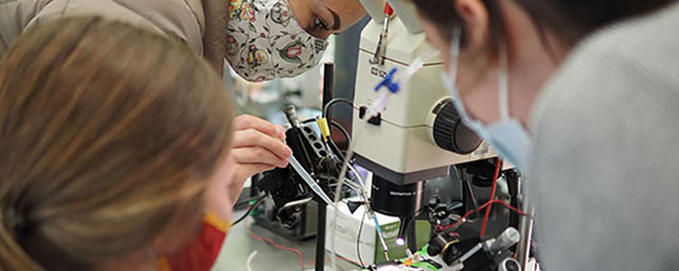
The Graduate Program in Neuroscience (GPN) at the University of Minnesota is a large interdisciplinary PhD program, made up of over 125 faculty members. Our goal is to provide training in neuroscience research across a broad range of techniques and disciplines, ranging from the molecular and genetic level to computational. Due to its interdisciplinary nature, our program is a highly collaborative and collegial environment in which to train.
Want to learn more about our program? Attend our virtual open house this fall!
Available dates and details can be found here

Education
We are an interdisciplinary program, with a productive and engaged faculty, committed to providing a supportive environment so that students can achieve their full potential.
Research
Our faculty are from 30 different departments and have a wide range of research interests and expertise. A complete list of faculty can be found here.
Community
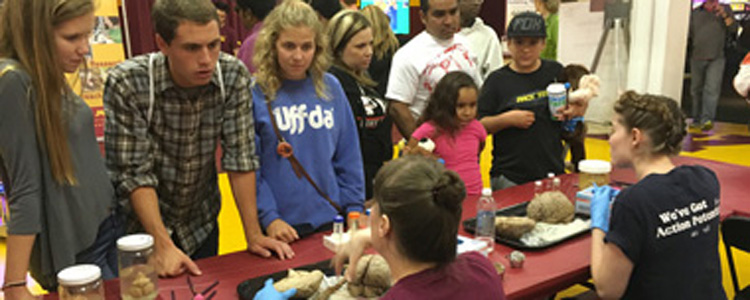
We support a number of programs that bring neuroscience into the community, such as Brain Awareness Week, Brains at the State Fair, and the Minnesota State Brain Bee.
Connect with us
 |
 |
 |